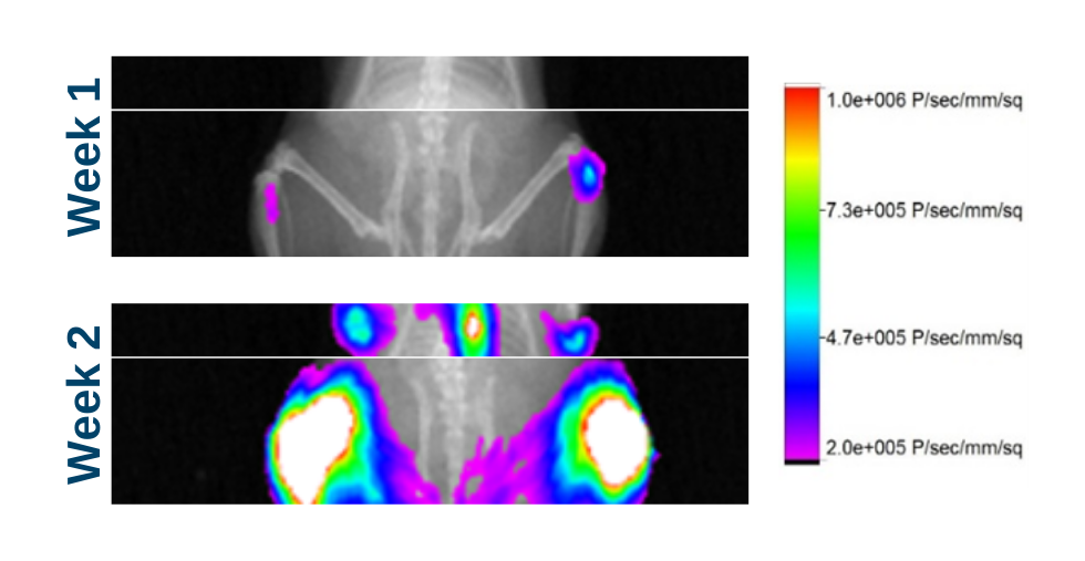
Leukaemic and Solid Tumour models are available. Luciferase labelled cells are monitored longitudinally using imaging to provide tumour progression data.
Orthotopic Models
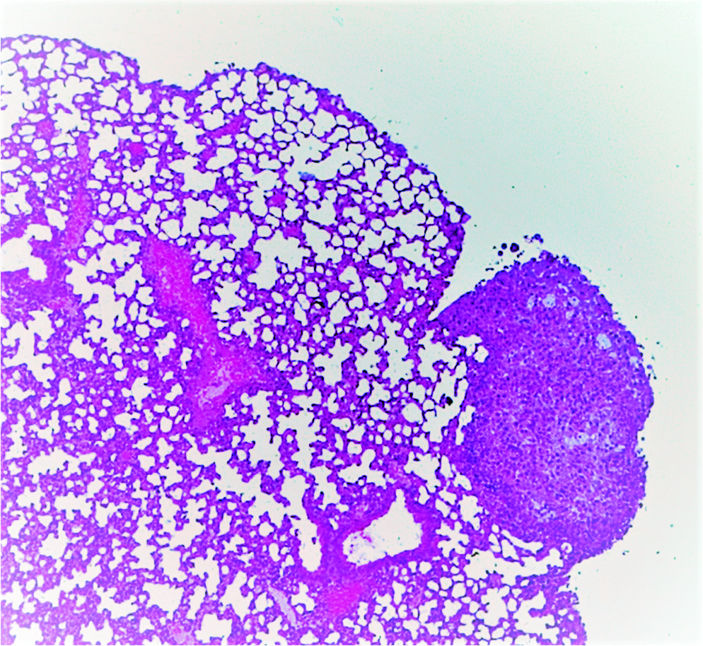
Cells are grown within the relevant body site, such as in the blood/bone marrow for leukaemia models (acute myeloid leukaemia and acute lymphocytic leukaemia) and the lung for lung cancer models.
Bespoke Services
In addition, we offer a bespoke service for new model development where we can luciferase-tag the cell line of your choice for future in vivo use.
Metastatic Models
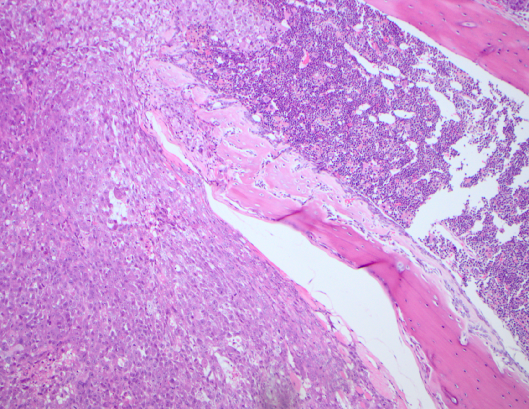
These models can be used to evaluate the efficacy of a drug on metastatic invasion and/or established metastatic lesions in secondary organs. Metastasis can occur via natural spread or be modelled by direct implantation into the metastatic site. For example, our ovarian cancer model is implanted into the intraperitoneal cavity, and the prostate cancer model can be implanted directly into the femur or intravenously to form liver metastases.


